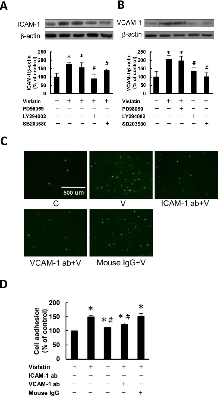
Fig. 2. Visfatin upregulates ICAM-1 and VCAM-1 expression in endothelial cells through the PI3K/Akt- and p38-dependent pathways. (A and B) Cells were pre-incubated for 1 h in the absence or presence of the ERK1/2 inhibitor PD98059 (PD, 30 μM), PI3K inhibitor LY294002 (LY, 30 μM), or p38 MAPK inhibitor SB203580 (SB, 20 μM), followed by incubation in the absence or presence of visfatin (100 ng/ml) in the continued absence or presence of the inhibitor for a further 24 h. The expression of ICAM-1 and VCAM-1 were measured. (C and D) Endothelial cells were left untreated or were incubated with 100 ng/ml visfatin alone or together with 10 mg/ml anti-ICAM-1, anti-VCAM-1 monoclonal antibodies, or 10 mg/ml mouse IgG for 24 h. The adhesion of THP-1 monocytes was measured. The results are the mean ± SEM for three separate experiments, each in triplicate. *P<0.05 compared with the untreated control; #P<0.05 compared with the visfatin alone group.